Cancer Research
Latest Research
Research Archive
- Research articles from 2025
- Research articles from 2024
- Research articles from 2023
- Research articles from 2022
- Research articles from 2021
- Research articles from 2020
- Research articles from 2019
- Research articles from 2018
- Research articles from 2017
- Research articles from 2016
- Research articles from 2015
- Research articles from 0
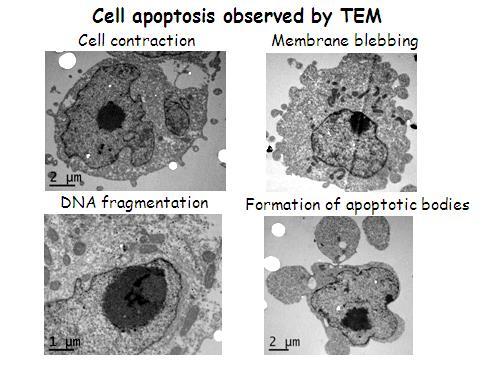
3-dimensional imaging of cancer cells by electron tomography

The University of Warwick
A number of metal-based compounds with promising anti-proliferative activity toward a wide range of tumours with novel mechanisms of action have recently been discovered. Such new metal-based anticancer drugs may be able to widen the spectrum of treatable cancers, reduce toxic side effects and overcome platinum resistance.
The Sadler group (Department of Chemistry, The University of Warwick) have been investigating the effect of novel organometallic iridium- and osmium-based potential anti-cancer compounds on various carcinoma cells by imaging fixed cell sections treated with the compound of interest (Liu et al., J. Med. Chem. 2011, 54, 3011-3026 and Van et al., Bioconjugate Chemistry. 2011, 22 (2), 218-226). Initial studies of sections of ovarian cancer cells have suggested that osmium anticancer compounds can cause significant changes in the morphology of cell organelles (see image). However – the images obtained contain ambiguities and it is often hard to identify precise structures in two dimensions.
Electron tomography offers a solution to this problem owing to it being an exceptionally powerful technique for obtaining 3 dimensional images of biological specimens. Better and clearer images of the architecture of essential components of these cells will provide key information on the effects of these anti-cancer compounds on both the morphology of cellular organelles and on cell function. In turn this will allow their potential as therapeutic drugs to be more effectively judged.
This project will compare the morphologies of non-resistant and resistant ovarian and colon cancer cells in the presence and absence of The Sadler group’s novel compounds. Imaging two types of cancer cells and comparing drug-resistant and drug-sensitive forms will allow the potential of this approach to be assessed for more extensive studies. It can be envisaged that the observation of the 3D morphological changes in cells could become a valuable anticancer drug screening assay.
The results of this study will be distributed widely to ensure that the wider community of scientists, clinicians and the pharmaceutical industry is aware of the potential of this technique.
(see also the News section for updates)