
News
In this section
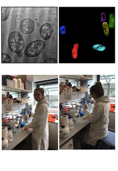
The Medical and Life Sciences Research Fund grant to Sarah Smith aimed to develop new electron microscopy techniques to image proteins in biological samples. The ultimate aim was to understand the changes that take place in cancer cells but Sarah first developed the techniques in simpler bacterial cells (E. coli). Most electron microscope techniques analyse samples in 2-dimensions, and a typical image of an E. coli cell is shown in the top left image. However, it is of great interest to understand the distribution of key proteins in 3 dimensions, and to do this Sarah worked with an electron microscope manufacturer, Jeol UK, who have developed the technique of 'array tomography' in which samples are taken from ultra-thin slices of the specimen and imaged in 2D, after which the images are assembled in 3D using new computer programmes. The location of specific proteins can be visualised by tagging them with gold-labeled antibodies. An example is shown in the top right hand image - in these samples a membrane protein was 'tagged' for visualisation and the images show that the membrane around the cells can be visualised with extremely high resolution. The system is now being used to track proteins in cancer cells and a manuscript on the E. coli work has been submitted for publication.
This 'proof-of-concept' work has succeeded in providing the preliminary data for a successful larger-scale grant application by Dr Corinne Smith. The grant, for £457,252, is funded by the Biotechnology and Biological Sciences Research Council (BBSRC).